Dentistry and oral surgery are some of the most common surgeries performed within the veterinary practice. Given the statistic that over 87 percent of dogs and 70 percent of cats will be affected by periodontal disease during their lifespan, add oral tumours, inflammatory conditions, malocclusions, tooth and jaw fractures and much more and it is not surprising that we spend much of our time in the dental suite (Enlund et al., 2020).
Managing anaesthesia for such cases can prove tricky and stressful. I imagine if we were to ask those regularly partaking in the management of dental and oral surgery and its associated anaesthesia what one of their most encountered concerns are, then preventing and treating hypothermia would be near, if not on top of, the list! It certainly is for me, so you are not alone in this challenge.
Inadvertent perioperative hypothermia is unfortunately common, with moderate to severe hypothermia developing in 32 percent of dogs and 70.9 percent of cats (Dugdale et al., 2020).
So, why do patients undergoing dental or oral surgery seem so prone to becoming hypothermic? What body temperature is classified as hypothermia? How can we prevent this? When it occurs, how can we treat it? Why do we need to treat it? This article aims to answer all these questions, and hopefully give you some ideas as to how we can improve our patient care regarding avoiding and treating hypothermia for patients undergoing anaesthesia for dental and oral surgery.
What is hypothermia and why do our patients experience it during dental and oral surgery?
Hypothermia in veterinary patients is classified as described in Table 1 (Brodeur et al., 2017). Although we should aim to keep our patients normothermic during all anaesthesia episodes, it is important to remember that what is normal for one patient may differ slightly for another. Obtaining a preoperative temperature is therefore paramount, so it is known what is deemed normal for each individual patient.
| Severity of hypothermia | Core body temperature |
|---|---|
| Mild | 36.7°C to 37.7°C |
| Moderate | 35.5°C to 36.7°C |
| Severe | 33°C to 35.5°C |
| Profound/critical | below 33°C |
Animals thermoregulate by mechanisms of convection, conduction, radiation and evaporation.
During any anaesthetic episode (not just for those undergoing dental surgery), the patient’s body temperature will likely decrease due to muscle inactivity, reduced metabolic rate, drug-induced peripheral vasodilation and depression of the thermoregulatory centre (Reiter and Gracis, 2018).
Although in dentistry we do not lose body temperature due to open cavities, nor by hair removal or the use of cleansing and antiseptic preparations which will wet the skin in preparation for surgery (Steagall et al., 2017), we do see body temperature drop for a variety of reasons, including:
- In smaller patients, a non-rebreathing anaesthesia circuit may be required; this will likely require a higher fresh gas flow, particularly if capnometry is not available. Oxygen is cold, and will chill the patients quickly when used at a higher rate
- Hypothermia is typically seen more frequently and rapidly in smaller patients due to their relatively large body surface area to volume ratio. Many of the patients presenting for oral surgery are cats or smaller-breed dogs
- Patients may not have been eating well prior to the surgery, and therefore have less fat and muscle to insulate them
- Depending on the surgeon’s preference for patient position during surgery, the patient may be turned regularly, causing body heat to be frequently released into the environment
- Copious amounts of cold water are used during oral surgery, for both the scaler and the drill; this is a necessity to avoid thermal burns but will quickly make for a wet and cold patient
- Many drugs that are used to facilitate sedation and anaesthesia will cause hypothermia by depressing the hypothalamic thermoregulatory centre (Steagall et al., 2017)
- Heat loss will start to occur as soon as the patient receives any sort of preanaesthetic medication (Reiter and Gracis, 2018)
- Cool environmental temperatures, both in the wards and in the surgical suite. Ideally the ambient temperature would be kept above 23°C to help prevent hypothermia
- Dentistry and oral surgery can take some time. The longer the duration of anaesthesia, the more likely the patient is to become hypothermic
- Currently, due to the pandemic and it being winter, many patients are having to wait in the car/kerbside. From this I have found that more patients are arriving with a slightly lower than normal temperature
Further risk factors for development of hypothermia under general anaesthesia include, but are not limited to: age (younger patients have immature thermoregulatory abilities and older patients may have less body fat and muscle coverage), poor body condition, hypothyroidism, higher ASA status, cardiac conditions and diabetes (Dugdale et al., 2020).
The importance of preventing hypothermia
Hypothermia brings with it a multitude of negative effects and outcomes for our patients (Bryant, 2010; Brodeur et al., 2017; Steagall et al., 2017), including:
- When a patient shivers, their oxygen requirements increase by 40 percent. Shivering is also not a pleasant experience, particularly if there are surgical wounds
- Recovery time will be prolonged, potentially by hours, if the patient returns to recovery hypothermic. This places the patient at risk and creates more work for the recovery team as the patient will need to be more closely monitored and nursed
- For every 1°C drop in body temperature, the mean alveolar concentration requirements (MAC) will reduce by 5 percent. This leaves the patient at risk of inadvertent overdose of the anaesthesia agent being used
- Anticholinergics will not be effective (such as atropine and glycopyrronium), thus making it harder to treat a hypotensive episode caused by reduced cardiac output originating from bradycardia
- Blood viscosity increases
- Pulse oximetry will not read as accurately in a hypothermic patient
- Arrhythmias due to slowed myocardial conduction, resulting in abnormal depolarisation and repolarisation, along with tachypnoea or respiratory depression as hypothermia progresses
- Splanchnic and hepatic perfusion decreases causing decreased hepatic metabolism and GI motility
- Delayed wound healing in the post-operative period
- Increased infection rates, causing wound breakdowns or revision surgery to be required
- For every 1°C drop in core temperature there is a 6 to 7 percent decline in cerebral blood flow, resulting in a concurrent deterioration in mentation
- Severe hypothermic cases or prolonged episodes of hypothermia can result in death of the patient
Preventing and treating hypothermia
Monitoring the patient’s temperature
The patient’s temperature must be monitored closely throughout the procedure, either continuously or at least every 15 minutes, sooner if there are marked changes. The temperature attained should be annotated on the anaesthesia form and action taken should the temperature increase or decrease; the action taken should also be noted (Bryant, 2010).
For dental and oral surgery, a rectal temperature is the easiest location to obtain a temperature from. Care should be taken that the thermometer is not placed into faeces, and that the thermometer is removed prior to recovery. Lubrication and a gentle technique should be used to prevent any structural damage.
Preventing hypothermia
All patients can suffer from hypothermia, so we must be pre-emptive in how we try to prevent this from occurring. Ideas to keep your patient warm include the following:
- Creating a warm environment, avoiding heavy use of air conditioning. An ambient temperature above 23°C is recommended, but may not be comfortable for staff members
- Placing a warm jacket or blanket on the patient from the time of premedication
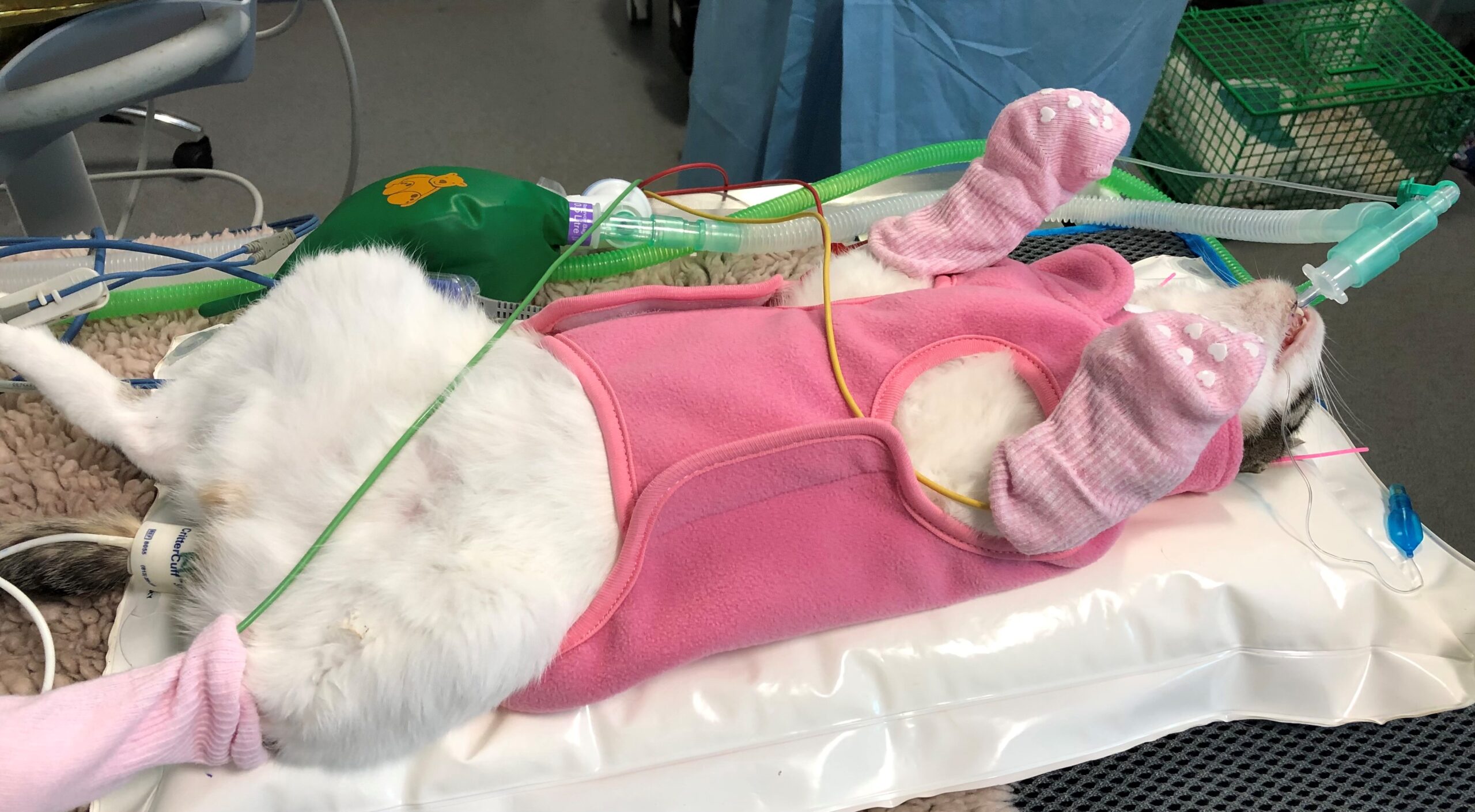
- Baby socks on paws
- Bubble wrap on extremities and around the skull
- Foil blanket around the body
- Plastic endotracheal tube ties; this prevents water from wicking up the tie which would create a wet skull area
- Soft absorbent mat for the head to lie on; this will drain water away from the patient’s head
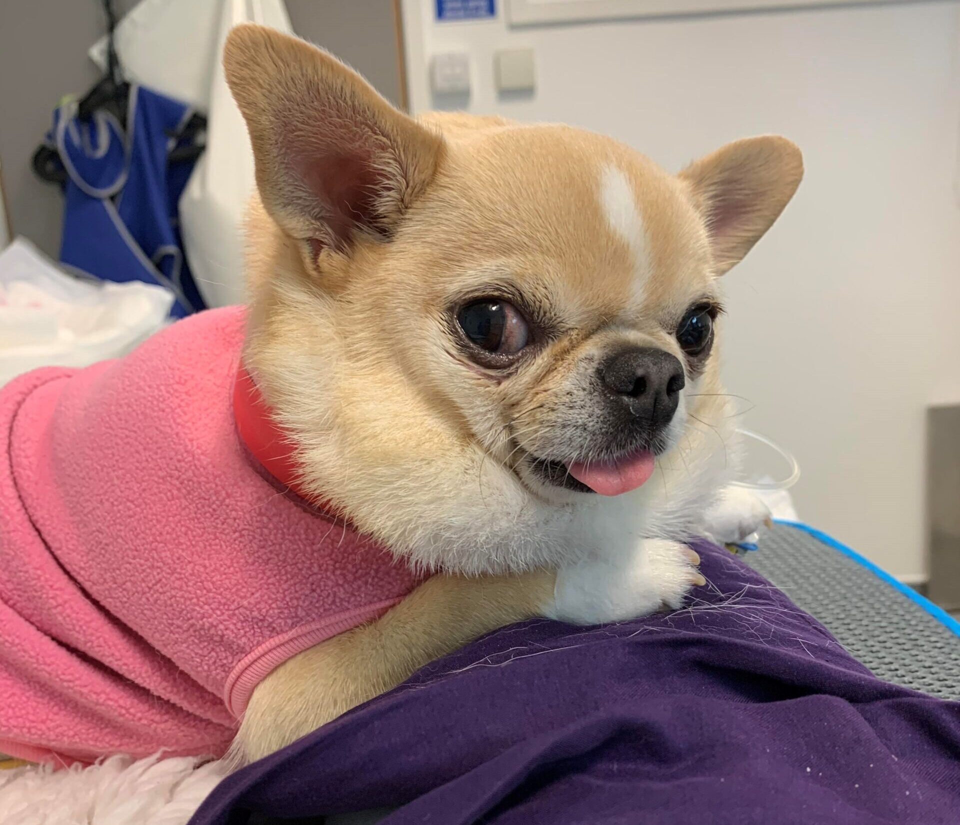
- Avoid turning the patient where possible. If the surgeon’s positioning means regular turning, the patient wearing a fleecy coat will prevent all the heat escaping each time the blankets are lifted to move the patient (Figure 1)
- Forced warm air blankets
- Hot water circulating mats
- Heat mats used with great care. The author only uses in patients under 5kg and using a thick vet bed between patient and mat
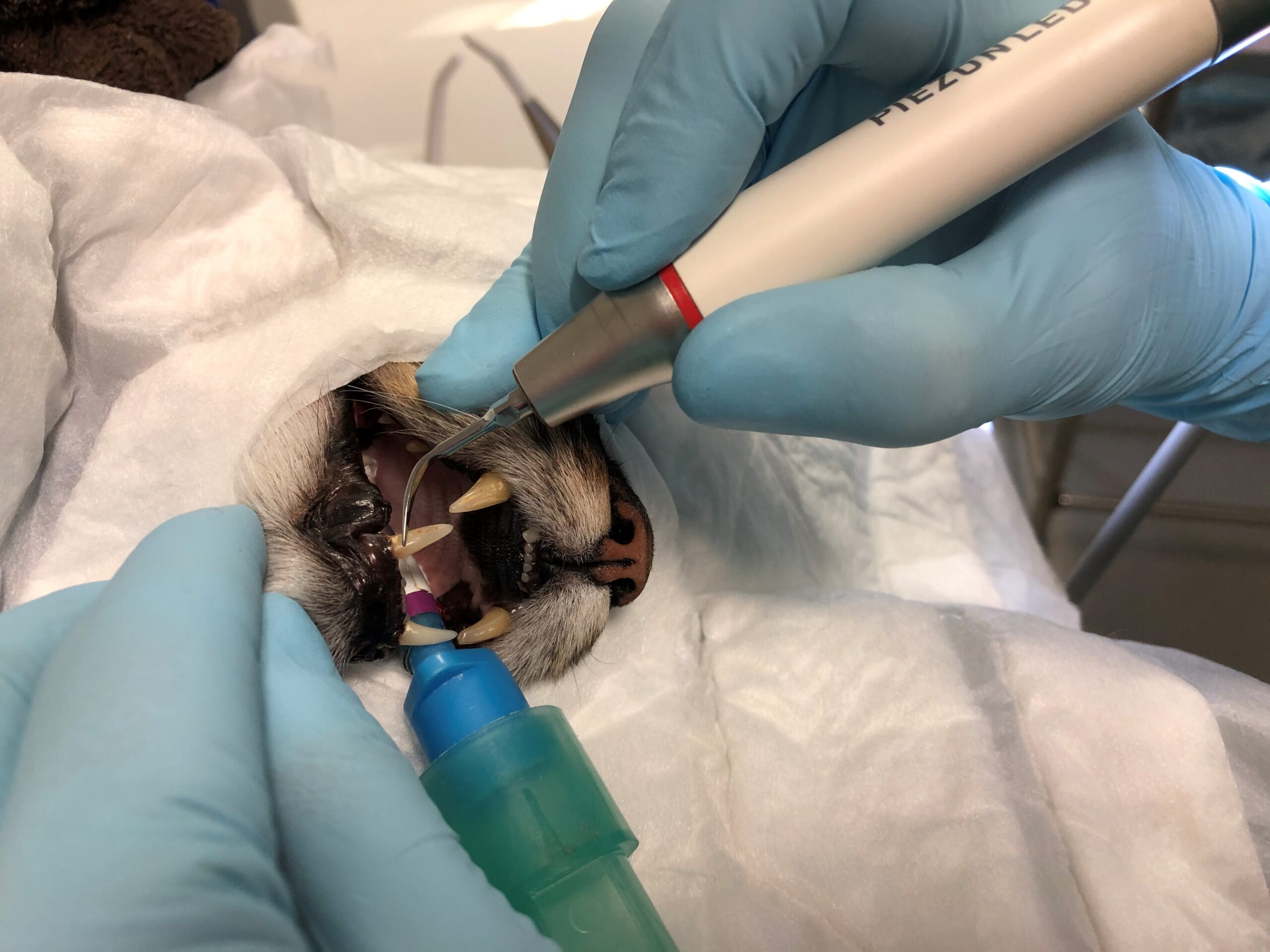
- Placing a puppy pad over the patient’s face to absorb all fluids. This will prevent the cold water from the dental drill and scaler from going into the patient’s fur. A hole is cut in the pad for the nose and mouth to go through (Figure 2). This will need to be changed as it becomes saturated
- Babygrows on smaller patients
- Cosy igloo beds for smaller patients pre- and post-op
- Use of fleece blankets over the patient to retain warmth
- Bean-filled beds to help insulate body heat; this will also maintain position and cushion any joints (Figure 3). Be careful not to mistake these with hot wheat-filled bags
- Heat moisture exchange, although this may get in the way of the surgeon
- Use of an incubator for recovery. This will also facilitate the administration of oxygen should it be required
- Fluid warmers; ideally intravenous fluids should be warmed to 38°C prior to being administered to patients – care must be taken not to overheat
Care should be taken if using:
|
Treating hypothermia
It is far better to prevent heat loss rather than facing the challenge of re-warming our patients. Caution must be taken when re-warming a patient to ensure it doesn’t happen too quickly. This may sound counterproductive, but if we warm a patient more than 1°C per hour it can cause tissue damage, with the vasodilation having the potential to cause an increase in intracranial pressure which will reduce cerebral perfusion and potentially worsen a neurological outcome (Dugdale et al., 2020).
Hyperthermia
Hyperthermia is equally as dangerous as hypothermia, although admittedly experienced less under general anaesthesia. Care should be taken with dogs who have very thick fur coats, or those who may have arrived in a very stressed condition as they will be more prone to hyperthermia.
Recovery
The close monitoring of the patient’s temperature should continue into the recovery period, with further warming aids used where needed. Kennels insulated with newspaper, kennel liners, thick bedding, blankets and a small pillow, or rolled-up blanket to simulate a pillow, can help keep the patient feeling secure and prevent further heat loss.
| Author’s top tips for keeping patients warm: 1) Fleecy jackets for all patients as soon as possible (Figure 1) 2) Covering the face with a puppy pad to prevent water from contacting the patient’s fur (Figure 2) 3) Socks on all paws as soon as under general anaesthesia and monitoring devices are in place (Figure 3) 4) Forced warm air blanket placed on the patient prior to experiencing hypothermia 5) The use of a plastic ETT tie prevents water from wicking up the tie and into the patient’s fur and muzzle area |
Summary
Hypothermia is detrimental to the health and welfare of our patients. We have established that prevention of hypothermia is superior to needing to increase a patient’s temperature and the RVN is perfectly placed to monitor and take all steps possible to prevent their patient from suffering the effects of hypothermia.
References (click to expand)
| Brodeur, A., Wright, A. and Cortes, Y. | 2017 | Hypothermia and targeted temperature management in cats and dogs. Journal of Veterinary Emergency and Critical Care, 27, 151-163 |
| Bryant, S. | 2010 | Anesthesia for Veterinary Technicians. Wiley Blackwell |
| Dugdale, A., Beaumont, G., Bradbrook, C. and Gurney, M. | 2020 | Veterinary Anaesthesia: Principles to Practice. Wiley-Blackwell |
| Enlund, K. B., Brunius, C., Hanson, J., Hagman, R., Höglund, O. V., Gustås, P. and Pettersson, A. | 2020 | Dog owners’ perspectives on canine dental health – a questionnaire study in Sweden. Frontiers in Veterinary Science, 7, 298 |
| Steagall, P., Robertson, S. and Taylor, P. | 2017 | Feline Anesthesia and Pain Management. John Wiley & Sons, Inc. |
| Reiter, A. M. and Gracis, M. | 2018 | BSAVA Manual of Canine and Feline Dentistry and Oral Surgery, 4th ed. British Small Animal Veterinary Association |