Glaucoma in cats remains a challenging condition to manage clinically, not least due to its often subtle presentation and cats’ notorious ability to mask pain and discomfort. Prompt recognition and appropriate intervention are essential to preserve vision and, in some cases, salvage the globe. This article will explore the pathophysiology, diagnosis and current management options for feline glaucoma and discuss considerations for referral.
By the end of this article, readers should be able to:
- Understand the underlying pathophysiology of feline glaucoma
- Recognise clinical signs and perform appropriate diagnostic tests
- Identify and initiate suitable medical and surgical treatment options where appropriate
- Evaluate when referral is indicated
- Consider quality of life and owner communication strategies in glaucoma cases
The pathophysiology of glaucoma
Glaucoma is defined as an increase in intraocular pressure (IOP) that exceeds the physiological tolerance of the eye, leading to optic nerve damage and progressive loss of vision. In the feline patient, it is typically a secondary condition rather than a primary disease process, most commonly associated with chronic uveitis, intraocular neoplasia, trauma or lens luxation.
In the feline patient, [glaucoma] is typically a secondary condition rather than a primary disease process, most commonly associated with chronic uveitis, intraocular neoplasia, trauma or lens luxation
Primary glaucoma is rare in cats and generally breed-specific, with Siamese, Burmese and Persian breeds reported to be at increased risk due to anatomical predispositions in the iridocorneal angle.
The underlying cause of raised IOP in most cases is impaired aqueous humour outflow. The conventional outflow pathway, via the iridocorneal angle and trabecular meshwork into the scleral venous plexus, becomes obstructed either physically (eg neoplasia, fibrin, inflammatory debris) or functionally (eg synechiae formation in chronic uveitis).
Clinical signs and diagnosis
Early diagnosis of feline glaucoma remains problematic. Cats may not present with overt pain or discomfort until IOP is markedly elevated. Subtle signs such as behavioural changes, reduced activity levels or hiding may be the only indicators.
Common clinical signs include:
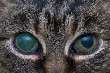
- Mydriasis, often unilateral initially
- Buphthalmos in more chronic cases
- Corneal oedema and vascularisation
- Haab’s striae (corneal striae from stretching of Descemet’s membrane)
- Blindness, either sudden or insidious
A comprehensive ophthalmic examination is critical and should include slit-lamp biomicroscopy, indirect ophthalmoscopy, tonometry and gonioscopy wherever possible
A comprehensive ophthalmic examination is critical and should include slit-lamp biomicroscopy, indirect ophthalmoscopy, tonometry and gonioscopy wherever possible. Tonometry provides an objective IOP measurement. Normal feline IOP ranges from 10 to 25mmHg; anything above 30mmHg is considered abnormal and warrants further investigation.
Imaging such as ocular ultrasound or CT/MRI may be helpful if neoplasia or posterior segment pathology is suspected. Electroretinography (ERG) may help determine visual potential in cases where the globe is structurally intact but vision is absent.
Management of glaucoma in cats
Management objectives
The primary goals of glaucoma management in feline patients are:
- To preserve vision, where possible
- To control IOP
- To alleviate ocular pain
- To identify and address the underlying cause
Management strategies can be broadly divided into medical therapy and surgical intervention. The choice of approach depends on the chronicity of the disease, visual status of the eye and presence of concurrent ocular pathology.
Medical management
In early or acute cases, particularly when vision is still present, medical management is the first line of treatment. Setting realistic expectations with owners is important, as the long-term prognosis for vision retention is guarded. In chronic or end-stage cases, medical management is often palliative. Depending on the clinical presentation, a range of pharmacological agents can be utilised:
- Topical carbonic anhydrase inhibitors: dorzolamide hydrochloride 2% is commonly used to reduce aqueous humour production via inhibition of carbonic anhydrase enzymes in the ciliary processes. This is often the first-line therapy in feline glaucoma due to its safety profile and efficacy
- Combination drops: a combination of dorzolamide 2 percent and timolol maleate 0.5 percent can be used for a synergistic effect. Timolol is a non-selective β-adrenergic antagonist that further suppresses aqueous humour production and may offer better IOP control than monotherapy
- Topical β-blockers: timolol may be used alone in early or mild cases, although it tends to be more effective when combined with a carbonic anhydrase inhibitor as mentioned above
- Prostaglandin analogues: latanoprost can be considered, but its efficacy in cats is limited due to reduced prostaglandin F receptor expression in the feline ciliary epithelium. In cases of secondary glaucoma associated with uveitis, it may worsen intraocular inflammation and is used cautiously, if at all
- Systemic carbonic anhydrase inhibitors: acetazolamide may be used in acute IOP crises, but is not typically recommended for long-term management due to systemic side effects, including metabolic acidosis and electrolyte imbalances such as hypokalaemia
- Topical anti-inflammatories: corticosteroids or NSAIDs can be used adjunctively where intraocular inflammation is present, provided corneal integrity is intact. These help reduce inflammatory components that contribute to increased IOP in secondary glaucoma
- Hyperosmotic agents: mannitol (IV) and oral glycerol can be used in emergency situations to achieve rapid, short-term IOP reduction by osmotically drawing fluid from the vitreous into the systemic circulation. These are particularly useful in acute glaucomatous crises but are contraindicated in patients with systemic dehydration, renal compromise or cardiovascular instability
Compliance can be a major limiting factor in feline patients, especially with frequent administration of eye drops. Transdermal and compounded options can be explored, but should be backed by pharmacokinetic data where possible.
Surgical management
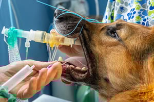
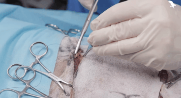
Surgical intervention in feline glaucoma is typically reserved for cases that are refractory to medical therapy or where the globe is irreversibly blind and painful
Surgical intervention in feline glaucoma is typically reserved for cases that are refractory to medical therapy or where the globe is irreversibly blind and painful. While the primary objective of surgery may be vision preservation, in most feline cases the goal is often palliative – relieving discomfort and improving quality of life. Available surgical interventions include:
- Evisceration with intrascleral prosthesis placement: this procedure involves the removal of intraocular contents while preserving the scleral shell and extraocular muscles. A silicone or acrylic prosthesis is placed within the sclera to maintain a more natural cosmetic appearance. It is suitable for patients with blind, non-infected, painful eyes, but should not be used where there is neoplasia or infectious uveitis
- Cyclodestructive procedures: these are aimed at reducing aqueous production by targeting the ciliary body, and may be considered in select vision-retaining cases or where globe salvage is desired:
- Transscleral diode laser cyclophotocoagulation (TSCP): this non-invasive technique uses infrared diode laser energy to ablate ciliary processes through the sclera. Its use in cats is technically challenging due to anatomical variability and a more fibrous sclera, and outcomes are variable
- Endoscopic cyclophotocoagulation (ECP): a more precise, albeit less commonly available, technique that directly visualises and ablates the ciliary processes from within the eye. It requires specialist equipment and anaesthetic stability, limiting its widespread use in general practice
- Anterior chamber shunt placement: glaucoma drainage implants (eg Ahmed or Molteno valves) are designed to facilitate aqueous outflow by creating an alternative drainage pathway to a subconjunctival bleb. These are rarely performed in cats due to a higher risk of fibrosis, shunt occlusion, hypotony and post-operative complications. However, they may be considered in select cases by experienced ophthalmic surgeons
- Enucleation: this remains the most definitive and commonly performed surgical treatment for end-stage glaucoma in cats. Removal of the globe provides immediate and permanent relief from intraocular pain, eliminates the need for ongoing medication and is often the treatment of choice when the eye is blind and cosmetically unsalvageable. It also facilitates histopathological assessment, which may be important in cases where underlying neoplasia is suspected
Alternatives such as evisceration and intrascleral prosthesis placement may be considered in select cases for cosmetic reasons, though are less common in feline practice due to globe size and the risk of complications.
Prognosis and follow-up
Owners should be educated about the progressive nature of the disease, the importance of compliance with medical therapy and the potential need for future surgical intervention or enucleation
The prognosis for visual preservation is, unfortunately, guarded in most feline glaucoma cases, particularly if secondary in nature. Early intervention and aggressive treatment are key to maintaining vision and comfort. Chronic cases often require lifelong management, and regular re-evaluation of IOP, vision and globe status is essential.
Owners should be educated about the progressive nature of the disease, the importance of compliance with medical therapy and the potential need for future surgical intervention or enucleation.
Referral considerations
Referral to an ophthalmologist may be considered when:
- The diagnosis is uncertain
- Vision is still present and specialist intervention may preserve it
- Intraocular neoplasia or lens luxation is suspected
- Surgical procedures such as cyclophotocoagulation (CPC) or shunt placement are indicated
Case studies
A 10-year-old domestic longhair cat
A 10-year-old neutered male domestic longhair cat was presented with a gradual onset of unilateral mydriasis and apparent vision loss in the left eye. The owner reported the cat had become more reclusive over the past week, preferring dark, quiet areas and avoiding jumping onto furniture.
On ophthalmic examination, the left eye exhibited mydriasis, mild corneal oedema and reduced menace response. IOP was 41mmHg OS (left eye) and 18mmHg OD (right eye). Fundic examination was limited due to corneal haze. The right eye appeared grossly normal. There was no history of trauma or recent illness.
The working diagnosis was unilateral secondary glaucoma, possibly due to previous undiagnosed uveitis. The cat was started on a topical dorzolamide/timolol combination twice daily, and systemic analgesia with buprenorphine was provided.
Within 48 hours, IOP remained elevated and vision was not restored. The decision was made to refer for advanced imaging and surgical planning. Enucleation was eventually performed due to globe discomfort and non-visual status.
A seven-year-old Siamese cat
A seven-year-old female spayed Siamese cat presented for a second opinion due to ongoing ocular discomfort and poor response to topical therapy. The referring vet had diagnosed anterior uveitis and prescribed topical steroids. There was no documented IOP measurement.
On presentation, the cat was noted to have marked buphthalmos, corneal neovascularisation and Haab’s striae of the left eye. The globe was firm on digital palpation. Tonometry revealed an IOP of 45mmHg OS and 20mmHg OD. The cat showed no dazzle or menace responses in the affected eye.
Further questioning revealed a history of trauma several months earlier, which had resolved uneventfully at the time.
Due to the chronicity and visual loss, enucleation was recommended. Histopathology confirmed post-traumatic glaucoma with angle closure and chronic fibrotic changes.
Summary
Feline glaucoma remains a complex and multifactorial condition, with management options dependent on the underlying cause, chronicity and visual status of the eye
Feline glaucoma remains a complex and multifactorial condition, with management options dependent on the underlying cause, chronicity and visual status of the eye. Early detection, targeted therapy and a pragmatic approach to pain management are crucial. While long-term visual prognosis is often poor, with timely intervention and client education quality of life can still be preserved.
For those looking to refine their skills in ophthalmic case management, the Improve International Ophthalmology programme offers an ideal opportunity to deepen your expertise and elevate the level of care you provide to your patients.
References (click to expand)
| Carter, J. | 2019 | Hypertensive ocular disease in cats: a guide to fundic lesions to facilitate early diagnosis. Journal of Feline Medicine and Surgery, 21, 207-219 |
| Hartley, C. | 2016 | Feline glaucoma: are you missing it? In: BSAVA Congress Proceedings 2016. British Small Animal Veterinary Association, Gloucester, pp. 166-167 |
| Komáromy, A. M., Koehl, K. L. and Park, S. A. _x000D_ _x000D_ | 2019 | The future of canine glaucoma therapy. Veterinary Ophthalmology, 22, 16-33 |
| McLellan, G. J. and Miller, P. E. _x000D_ _x000D_ | 2011 | Feline glaucoma: a comprehensive review. Veterinary Ophthalmology, 14, 15-29 |
| Ricci, C. L., Passareli, J. V. G. C., Nascimento, F. F., Kanashiro, G. P., da Costa Zulim, L. F., Giuffrida, R. and Andrade, S. F. | 2024 | Comparison of three methodologies for measuring intraocular pressure in healthy cats. Veterinary World, 17, 1803-1809 |
| Tuleski, G. L. R., Pscheidt, M. J. G. R., dos Santos, J. P. and Sousa, M. G. | 2022 | Timolol 0.5% ophthalmic solution influences cardiac function in healthy cats. Journal of Feline Medicine and Surgery, 24, 1153-1160 |
| Webb, T. E. R. _x000D_ _x000D_ | 2021 | A review of glaucoma surgical therapy. Veterinary Ophthalmology, 24, 34-38 |
| Whelan, N. _x000D_ | 2021 | Treatment of glaucoma in animals. In: MSD Veterinary Manual. Merck and Co., New Jersey [Accessed: June 2025] |
| Woodham-Davies, S. | 2021 | The management of glaucoma in cats and dogs to include medical and surgical intervention. Veterinary Nursing Journal, 36, 350-353 |